Two large veins deliver oxygen-poor blood to your right atrium. All products are produced on-demand and shipped worldwide within 2 - 3 business days.

Heart Anatomy Anatomy And Physiology Ii
View Lab 5 Heart Anatomydocx from BIOLOGY 1280 at Hibbing Community College.

. 69055 Add to Lightbox. Heart Anatomy Self Test. Heart Posterior View Variant Image ID.
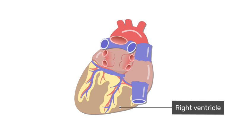
The transparent setting of the heart 3D model makes it easier to see the. The external anatomy of the heart has been set to Transparent but could easily be changed to Show. With the 3D model a student can tilt and rotate the heart to get an optimal view of each coronary vessel.
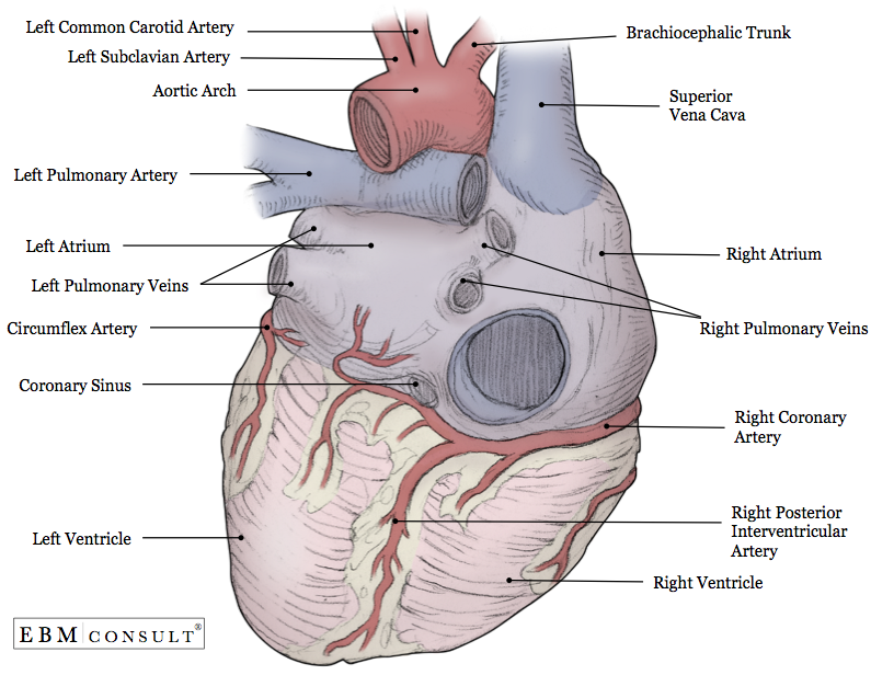
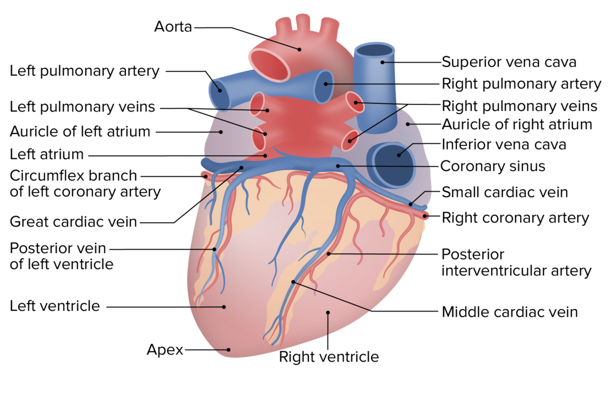
Ascending aorta Superior vena cava Right pulmonary artery Right pulmonary veins Right atrium Inferior vena cava Auricle of right atrium. The heart functions by pumping blood both to the lungs and to the systems of the body. In this image you will find superior vena cava right pulmonary artery right pulmonary veins right atrium inferior vena cava coronary sinus right coronary artery coronary sulcus posterior interventricular artery in it.
Looking for design inspiration. The photograph may be purchased as wall art home decor apparel phone cases greeting cards and more. 283 PLATE 4 EXPLANATION OF FIGURES 7 Internal bundles of the right atrium of the human heart posterior viewThe vena cavae have been opened through their posterior walls.
The atrioventricular AV valves. Anatomy and Physiology Posterior structures of the heart Label the following structures in the posterior view of the heart. This quiz has tags.
Atrioventricular and semilunar valves. The LMCA passes behind the right ventricular outflow tract and may extend for 0-10mm. Pumps deoxygenated blood to the lungs.
Anterior View Of Human Heart Anatomy is a photograph by Alayna Guza which was uploaded on September 7th 2017. Click on the tags below to find other quizzes on the same subject. There is a printable worksheet available for download here so you can take the quiz with pen and paper.
This is an online quiz called Label the Heart Posterior View. Images on Similar Topics. Heart Posterior View Variant Image ID.
The left ventricle is the major pumping chamber on the lower left side of the heart that ejects blood into the systemic circuit via the aorta and receives blood from the left atrium. Carry deoxygenated blood out of the right ventricle and into the lungs. To help simplify things we can convert the heart into a square.
Posterior View When you point to any structure on the photograph that region or structure will be highlighted in the smaller image to the left to help you locate it. Enlarged vein from junctions of coronary veins which empty. SLIDE 4 This is a posterior view of the heart.
There is a printable worksheet available for download here so you can take the quiz with pen and paper. Posterior View Choose a structure from the pull-down list. The right ventricle is the major pumping chamber on the lower right side of the heart that ejects.
An answer will then appear in the small window. Posterior View Of Heart Anatomy. Pumps oxygenated blood to the body.
The inferiorvena cava is spread widely open. Link this page. There are 4 chambers labeled 1-4 on the diagram below.
To prevent blood from flowing backwards or regurgitating back into the heart a system of one-way valves are present in the heart. Link this page. Up to 10 cash back The American journal of anatomy.
Aortic arch Left pulmonary artery Left pulmonary veins Auricle of left atrium Left atrium Circumflex artery in atrioventricular sulcus Coronary sinus Left ventricle c Posterior view of the external heart 2019 Pearson Education Inc. Receives deoxygenated blood from the body. Heart Anatomy Using your OpenStax text sections 191-192.
20864 Add to Lightbox. Enlarged vessel on the posterior aspect of the heart that empties blood into the right atrium. Your heart is divided into four chambers.
The boxes are numbered to correlate with the labeled chambers on the cartoon diagram. The LMCA arises from the upper portion of the left sinusjust below the sinotubular ridge of the aorta. LEFT CORONARY ARTERY ANATOMY.
This view allows a student to observe the major coronary vessels of the heart. The diameter of the LMCA ranges from 3-6mm. You have two chambers on the top atrium plural atria and two on the bottom ventricles one on each side of the heart.
Then click on the matching structure in the large heart image. Descending aorta pulmonary veins. One of these vessels the coronary sinus is returning to the right atrium the blood that has been to heart muscle SLIDES The next few slides focus on the coronary blood vessels.
If you click your left mouse button the name of that structure will appear to identify it. We will then divide that square into 4 different boxes which will represent the 4 chambers of the heart. Largest artery in the body.
One of the primary pumping chambers of the heart located in the lower portion of the heart. The heart valves can be broken down into two types. You may also find posterior interventricular sulcus middle cardiac vein right ventricle apex apex of heart left ventricle.
Runs alongside the anterior interventricular artery. This Illustration was published in. This is an online quiz called Anatomy of the Human Heart - Posterior View.
Label the 4 chambers as well as the major vessels entering and leaving these chambers.
Posterior View Of The Heart Diagram Quizlet

Posterior View Of The Heart Heart Anatomy Heart Diagram Anatomy

Posterior View Of The External Heart Diagram Quizlet
Anatomy Heart External

4 Posterior View Of The Human Heart Download Scientific Diagram

Heart Anatomy Labelled Illustration Stock Image C043 4821 Science Photo Library
The Heart Chambers And Their Functions
Heart Anatomy Concise Medical Knowledge
0 komentar
Posting Komentar